

Researchers at Rice University have developed spinalNET, a new implantable sensor that can record the electrical activity of spinal neurons in freely moving objects. This breakthrough could help unravel the complexity of how spinal neurons process sensory and motor functions, potentially leading to better treatment options for spinal cord diseases and injuries.
Implantable technologies have greatly improved our ability to study and even modulate the activity of neurons in the brain. However, neurons in the spinal cord are more difficult to study in action.
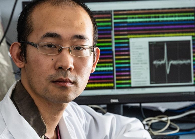
“If we understood exactly how neurons in the spinal cord process sensations and control movement, we could develop better treatments for spinal cord diseases and injuries,” says Yu Wu, a scientist who is part of a team of neuroengineers at Rice University working to solve the problem.
Introducing spinalNET: A breakthrough in neural monitoring
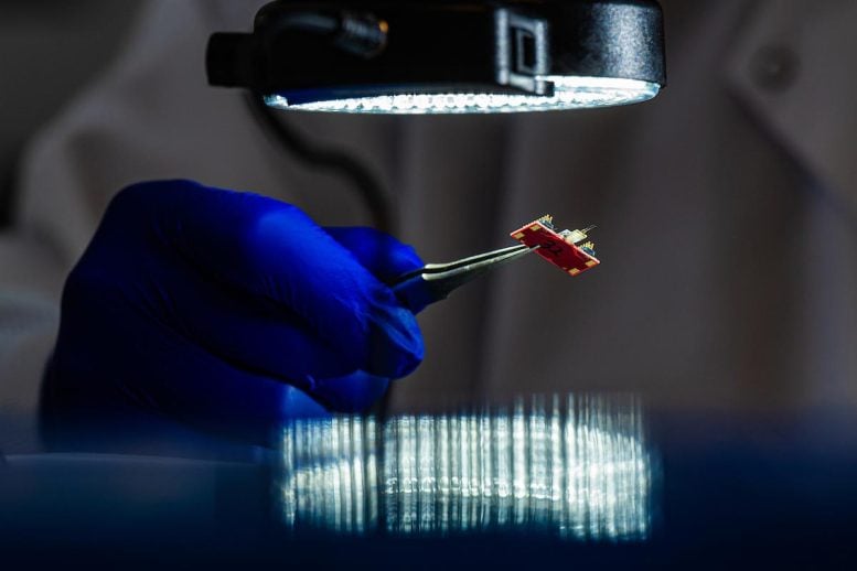
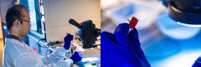
“We have developed a tiny sensor called spinalNET that records the electrical activity of spinal neurons while the subject performs normal activities without any restrictions,” said Wu, the lead author of a study on the sensor published in Cell reports. “The ability to gain such insights is a first but important step toward developing cures for the millions of people suffering from spinal cord diseases.”
According to the study, the sensor recorded neuronal activity in the spinal cord of freely moving mice over long periods of time and at high resolution, even tracking the same neuron over several days.
Challenges of spinal cord monitoring
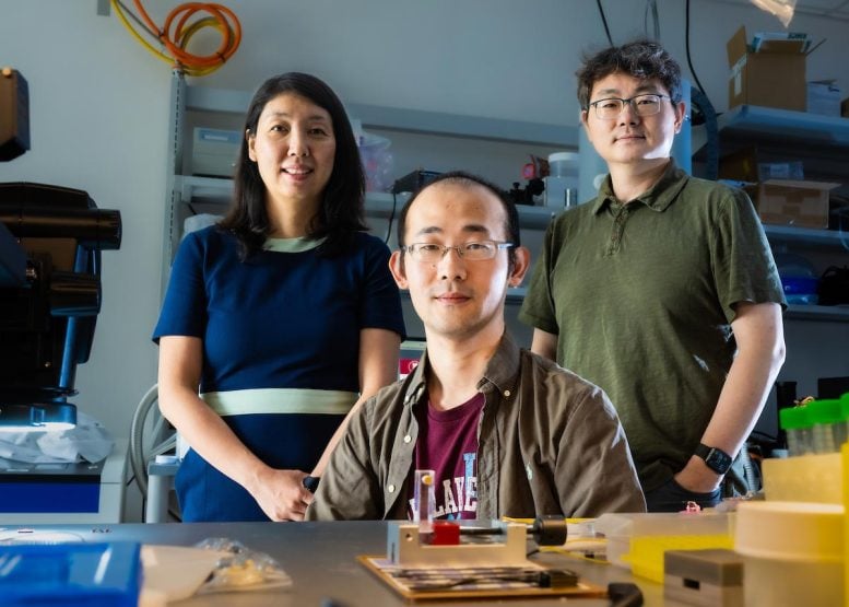
“Until now, the spinal cord has been more or less a black box,” said Lan Luan, associate professor of electrical and computer engineering and corresponding author of the study. “The problem is that the spinal cord moves so much during normal activity. Every time you turn your head or bend over, the spinal cord neurons move too.”
During such movements, rigid sensors implanted in the spinal cord inevitably disturb or damage the sensitive tissue. SpinalNET, on the other hand, is over a hundred times smaller than a hair and is therefore extremely soft and flexible – almost as soft as the nerve tissue itself.
“This flexibility gives it the stability and biocompatibility we need to safely record spinal neurons during spinal cord movements,” said Chong Xie, associate professor of electrical and computer engineering and of biological engineering and corresponding author of the study. “With spinalNET, we were able to obtain low-noise signals from hundreds of neurons.”
Findings and future directions in spine research
The spinal cord plays a critical role in controlling movement and other vital functions. The ability to record spinal neurons during unconstrained movement with fine spatial and temporal resolution provides insight into the mechanisms that make this possible. Using spinalNET, researchers discovered that the spinal neurons in the central pattern generator – the neural circuit that can generate rhythmic movement patterns such as walking without specific timing information – appear to be involved in much more than just rhythmic movement.
“Some of them are strongly correlated with leg movement, but surprisingly, many neurons have no obvious correlation with movement,” Wu said. “This suggests that the spinal circuit controlling rhythmic movement is more complicated than we thought.”
The researchers said they hope to unravel some of that complexity in future research, looking, for example, at differences in the way spinal neurons process reflex movements – such as startle – compared with voluntary actions.
“In addition to the scientific evidence, we believe that with further development, the technology has great potential as a medical device for people with neurological diseases and spinal cord injuries,” said Luan.
Reference: “Ultraflexible electrodes for recording neuronal activity in the spinal cord of mice during motor behavior” by Yu Wu, Benjamin A. Temple, Nicole Sevilla, Jiaao Zhang, Hanlin Zhu, Pavlo Zolotavin, Yifu Jin, Daniela Duarte, Elischa Sanders, Eiman Azim, Axel Nimmerjahn, Samuel L. Pfaff, Lan Luan and Chong Xie, May 9, 2024, Cell reports.
DOI: 10.1016/j.celrep.2024.114199
Funding: The research was supported by the National Institutes of Health (R01NS102917, U01NS115588, U01NS131086, R01NS109361, R01NS123160, R01NS108034, U19NS112959), Rice, the Salk Institute, and the Mary K. Chapman Foundation.
